

RSNA 2026
From November 29 to December 3, 2026, Vermon will be at RSNA in Chicago, Booth 8044. Come and discuss clinical use cases and discover our ultrasound transducer solutions.
Chicago, USA
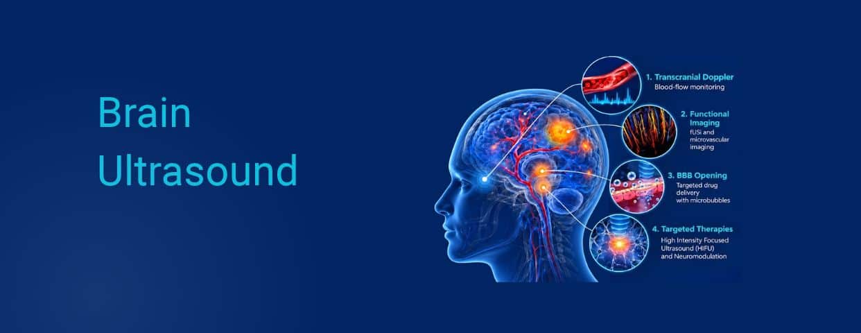
For many years, the adult human brain remained one of the most difficult organs to explore using conventional ultrasound methods. The reason is largely physiological: the skull creates a complex acoustic barrier, attenuating and distorting the propagation of ultrasound waves. As a result, MRI and CT became the reference modalities for brain imaging.
But advances in transducer architecture, aberration correction, acoustic focusing, ultrafast acquisition, and signal processing are gradually changing the landscape.
Today, ultrasound is no longer limited to studying cerebral blood flow. It can help map the microvasculature, indirectly observe functional brain activity, and, in specific configurations, interact with brain tissue. This evolution takes brain ultrasound beyond imaging and into an entirely new range of applications.
Transcranial Doppler, or TCD, is one of the earliest applications of ultrasound in the brain. Using relatively low frequencies, typically around 2 MHz, this modality measures the velocity and characteristics of blood flow in major cerebral arteries through specific acoustic windows in the skull.
It is used, among other applications, to:
– Monitor vasospasm following subarachnoid hemorrhage
– Assess certain arterial stenoses or occlusions
– Detect embolic signals
– Contribute to the evaluation of cerebral circulatory arrest
A major advantage is its ability to deliver continuous, real-time hemodynamic insights right at the patient’s bedside.
While conventional TCD uses a single-element probe positioned “blindly,” transcranial color-coded duplex sonography (TCCS) relies on compact, low-frequency phased arrays that image the vessels while measuring their flow. Their small footprint fits the narrow temporal acoustic window, and their sector-shaped field of view covers the main arteries of the circle of Willis. For example, Vermon’s 2.75 MHz, 96-element single-crystal phased array was built for this use.
Current development projects aim to make these examinations more reproducible and less operator-dependent through automated positioning, robotics, and advanced data processing.
The emergence of ultrafast imaging and advanced signal-processing techniques has opened the way to ultrasound super-resolution imaging, also known as Ultrasound Localization Microscopy, or ULM.
The principle relies on injecting contrast microbubbles into the bloodstream. By combining ultrafast imaging with precise tracking of individual microbubbles, researchers can reconstruct an extremely detailed map of the vascular network.
Studies in patients have demonstrated the ability to visualize transcranial brain vasculature at resolutions on the order of a few tens of micrometers, while also analyzing highly detailed blood-flow dynamics. In one patient, the technique even made it possible to visualize complex flow patterns such as vortices within a small, deep-seated cerebral aneurysm.
This represents a major shift in scale: ultrasound is no longer limited to major blood vessels but can begin to provide access to cerebral microcirculation. Although the technique is still largely confined to research settings, it showcases the extraordinary evolution of ultrasound imaging.
Another major development is Functional Ultrasound Imaging, or fUSi. Unlike conventional anatomical ultrasound, the objective is not simply to visualize brain structures.
Using ultrafast imaging, fUSi can detect very small variations in blood flow and blood volume within the cerebral microvasculature. Because these hemodynamic changes are associated with brain activity, they can be used to map the activation of different brain regions.
Researchers can therefore study functional connectivity, responses to different stimuli, or the impact of pharmaceutical compounds through applications such as pharmaco-fUSi. Proof-of-concept studies have already been carried out in humans, including intraoperative functional imaging, although the technology is still primarily used in research environments.
At the same time, volumetric imaging approaches such as matrix-array or row-column array (RCA) architectures are paving the way toward 3D and 4D ultrasound imaging, enabling the exploration of entire volumes rather than a single ultrasound plane.
This shift from anatomical imaging toward vascular, functional, and volumetric information is one of the most promising developments in brain ultrasound.
The paradigm shift becomes even more significant when ultrasound moves from imaging to actively targeting brain tissue.
HIFU is one of the most mature examples. By concentrating acoustic energy onto a very precise target, ultrasound can generate a localized, non-invasive rise in temperature sufficient to produce tissue ablation.
In the United States, the FDA approved an MR-guided focused ultrasound system in 2016 for selected patients with medication-resistant essential tremor. Indications were later expanded to include certain manifestations of Parkinson’s disease. In July 2025, approval was extended to staged bilateral treatment in advanced Parkinson’s disease.
Thermal ablation of brain tumors, including glioblastoma, remains investigational: skull heating and a treatment envelope limited to deep, central targets restrict its use, and most neuro-oncology efforts now focus on the lower-intensity approaches described below.
Another particularly active field of research involves the Blood-Brain Barrier, or BBB. While this natural barrier is essential for protecting the brain from neurotoxins and pathogens, it represents a major obstacle to targeted drug delivery. By restricting the passage of nearly all systemic therapeutics, it severely limits the ability to treat neurodegenerative diseases and brain tumors.
One approach under investigation combines low-intensity focused ultrasound with circulating microbubbles. Under the effect of the acoustic field, the microbubbles oscillate within the microvasculature of the targeted area. This mechanical interaction can temporarily and locally increase the permeability of the Blood-Brain Barrier, allowing specific therapeutic agents to reach brain tissue more effectively.
The effect is temporary, and the barrier subsequently returns to its initial state. This technology is currently being investigated in several clinical research areas, including neuro-oncology and neurodegenerative diseases.
At even lower acoustic intensities, ultrasound opens another pathway: ultrasound neuromodulation. Here, the objective is to influence the activity of targeted neural networks.
Research into LIFU (Low-Intensity Focused Ultrasound) and LIPU (Low-Intensity Pulsed Ultrasound) is exploring the possibility of activating or inhibiting neuronal activity non-invasively and reversibly. Research is currently underway in areas such as treatment-resistant depression, epilepsy, and a range of neurological and psychiatric disorders.
Upcoming developments could go even further. Research is already exploring wearable transducers, ergonomic ultrasound architectures, systems capable of combining transmission and reception, and the integration of functional imaging with neuromodulation. This convergence is also contributing to the development of non-invasive brain-computer interfaces.
Ultrasound may therefore progressively evolve from a technology used to measure blood flow to one capable of visualizing microvascularization, observing brain function, targeting specific areas, and potentially interacting with neural activity.
Despite their diversity, all these applications share one thing in common: they impose very different acoustic and engineering requirements. Passing through the skull, achieving sufficient sensitivity to detect low-flow signals, generating ultrafast images, precisely focusing acoustic energy, controlling output power, or miniaturizing a matrix-array architecture do not require the same type of transducer.
Frequency, geometry, element number and distribution, piezoelectric materials, electronic architecture, thermal management, and mechanical integration all become critical design parameters. This ability to engineer the ultrasound solution around the application is one of the central technological challenges in the future of brain ultrasound.
At Vermon, this evolution is directly connected to our core expertise: developing advanced transducer technologies capable of supporting the next generation of ultrasound applications, from imaging to targeted interaction with the brain.

RSNA 2026
From November 29 to December 3, 2026, Vermon will be at RSNA in Chicago, Booth 8044. Come and discuss clinical use cases and discover our ultrasound transducer solutions.
Chicago, USA
Read More

MEDICA 2026
Join Vermon at MEDICA 2026 in Düsseldorf to discover our custom and standard ultrasound probe solutions.
Düsseldorf, GER
Read More

IEEE IUS 2026
Join Vermon at IEEE IUS 2026 in Raleigh, NC (Oct 4–8). Visit us at booth 40 & 41 to discuss custom and standard ultrasound transducer solutions.
Raleigh, North Carolina
Read More

News Vermon Group
Vermon launches Vermon Group to accelerate its growth, combining the acquisition of Moduleus, expanded U.S. manufacturing capacity, and a unified platform for ultrasound solutions.
Read More

WHX 2026
Join Vermon at WHX 2026, in Dubai, UAE, from February 9 to February 12, North Hall 21, K.92.
Dubai, UAE
Read More