

Cardiovascular
Cardiovascular diseases are the top cause of death worldwide. Early detection of these conditions enables practitioners to diagnose and treat patients effectively.
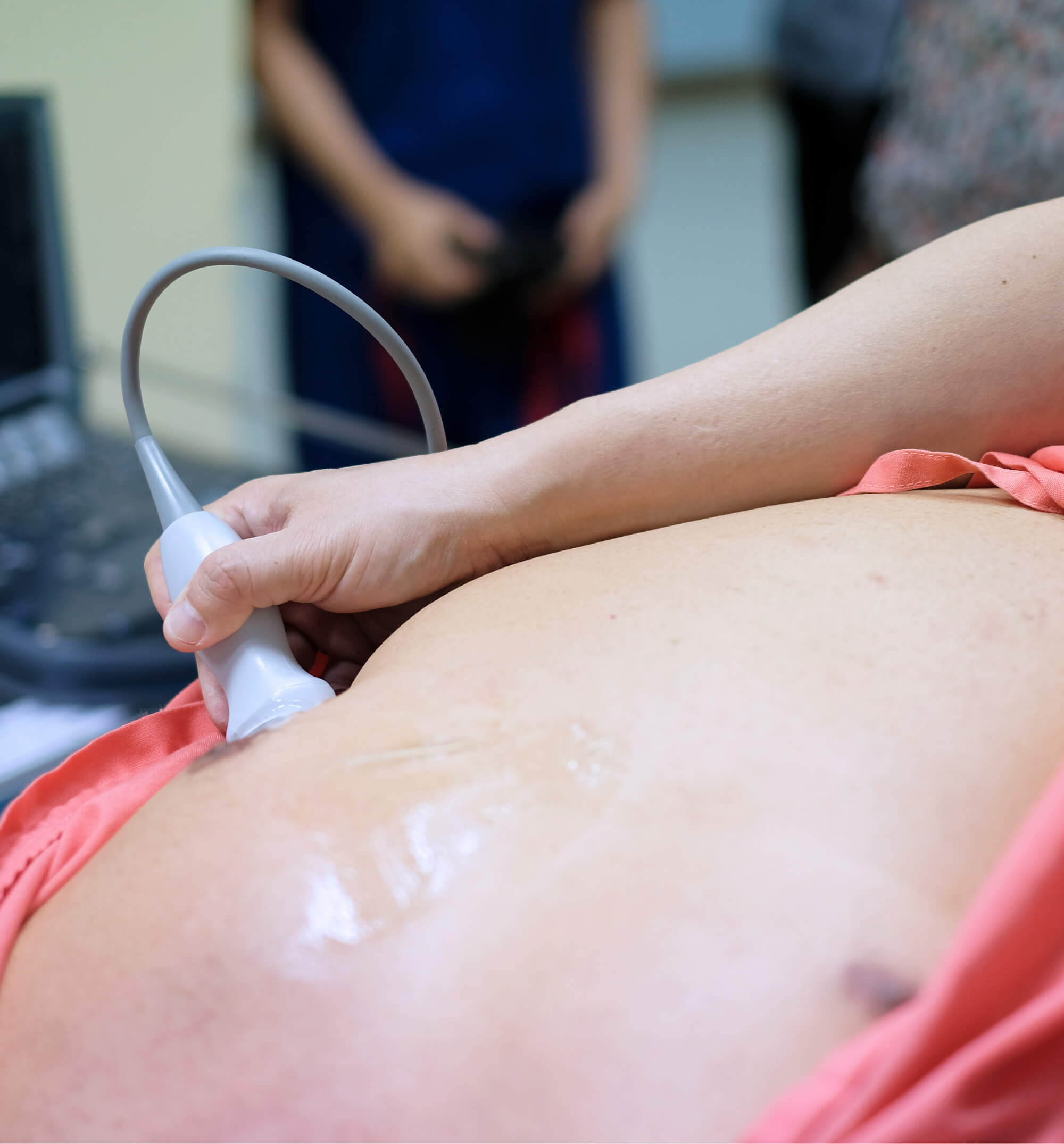
Cardiovascular ultrasound offers significant benefits such as portability (over MRI and CT scans), immediate availability, superior temporal resolution, quick examination times, and simplicity in performing clinical assessments.
Heart
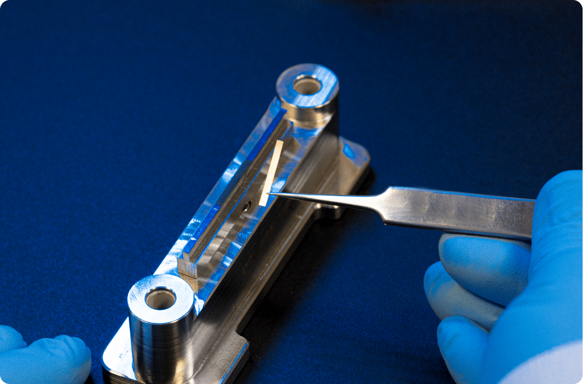
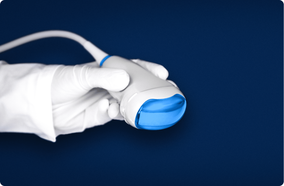
Known as echocardiography, the ultrasound investigation of the heart is performed using a small footprint phased array probe, typically operating at around 3.0 MHz.
One of the major aspirations of cardiologists is to be capable of identifying the morphology, dynamics, and kinetics of the complete heart structure throughout one entire heartbeat.
Transthoracic Echocardiography (TTE) represents the most often used imaging modality, providing invaluable and non-invasive information for heart diagnosis.
At each probe position, angulation and rotation, different views of the heart are accessible to provide tomographic images of cardiac structures and blood flow. Through the apical window and the subcostal window, TTE obtains relevant information and quantification of cardiac and myocardial morphology.
Cardiac function and perfusion; wall motion and deformation; valve function; and hemodynamics are studied using visual assessment, myocardial strain imaging, or Doppler blood flow quantification.
1
Heart
Recommanded probes :
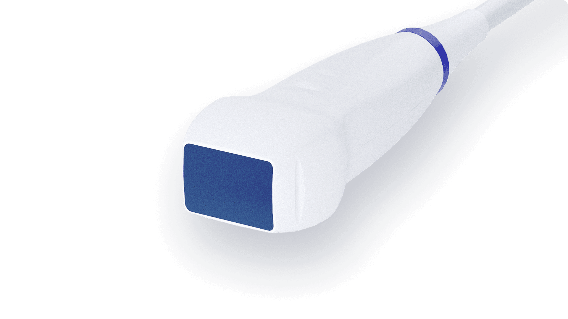
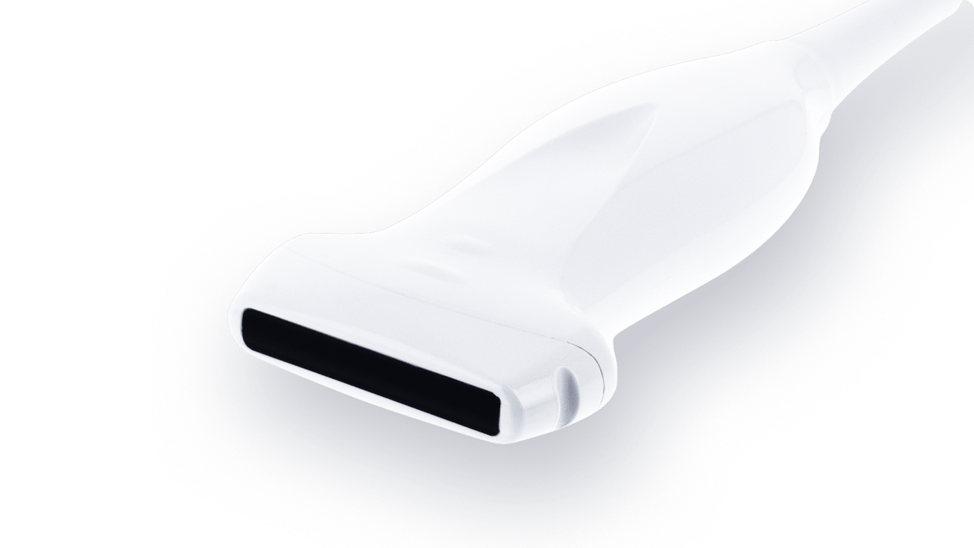
Phased Array – 2.8MHz – 96 elts
PA 2.8/96-SiX
-

Center Frequency 2.8
-

Number elements 96
-

Pitch 0.2
-

Radius curvature N/A
-

Transverse aperture 12
-

Focal Depth 80
Phased Array – 3.2MHz – 64 elts
PA 3.2/64-SiX
-
Center Frequency 3.2
-
Number elements 64
-
Pitch 0.24
-
Radius curvature N/A
-
Transverse aperture 12
-
Focal Depth 80
Brain
Brain ultrasound is usually carried out with a phased array probe through the temporal bone, typically operating at central frequencies from 2.5 MHz to 3.5 MHz.
Transcranial Doppler (TCD) ultrasound provides non-invasive and real-time measures of the cerebrovascular function assessed with Doppler blood flow. TCD is used to measure blood flow velocities in the basal artery and the Middle Cerebral Artery (MCA), terminal branch of the internal carotid artery.
TCD is a useful tool to detect embolic signals, the principal cause of cerebrovascular disorders. Acute ischemic stroke is characterized by a sudden loss of blood circulation resulting in a loss of the corresponding area neurologic function.
2
Brain
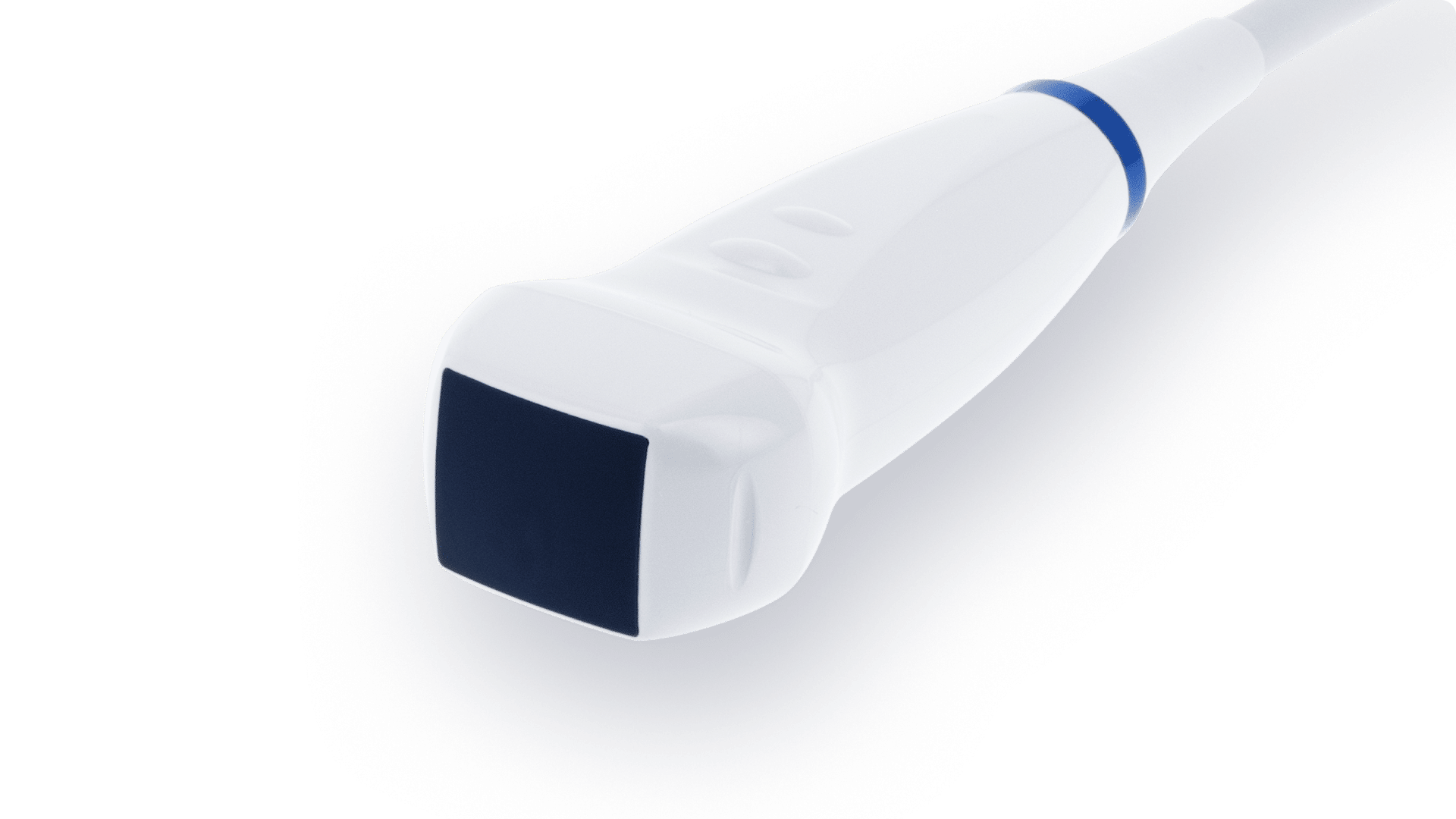
Recommanded probes :
Phased Array – 2.8MHz – 96 elts
PA 2.8/96-SiX
-
Center Frequency 2.8
-
Number elements 96
-
Pitch 0.2
-
Radius curvature N/A
-
Transverse aperture 12
-
Focal Depth 80
Phased Array – 2.8MHz – 80 elts
PA 2.8/80-SiX
-
Center Frequency 2.8
-
Number elements 80
-
Pitch 0.24
-
Radius curvature N/A
-
Transverse aperture 14
-
Focal Depth 80
Peripheral Artery & Veins
For ultrasound examinations of lower limbs’ venous and arterial system, a linear array probe with a frequency of around 6.0 MHz is usually appropriate. For the upper limbs, a linear array probe with a frequency higher than 7.5 MHz is recommended.
Ultrasound is the ideal modality for the assessment of the location and extent of peripheral vascular diseases because it is cost effective, reproducible, painless and non-invasive.
As a complementary tool for diagnosing abnormal blood flow, non-imaging CW Doppler may be used as a reference system to detect segmental systolic blood pressure for Ankle-Brachial Pressure Index (ABPI) examination.
Upper limb studies tend to be carried out less often than lower limb studies, because of the lower prevalence of atherosclerosis.
3
Peripheral Artery & Veins
Recommanded probes :
Linear Array – 6.0MHz – 192 elts
LA 6.0/192-SiX
-
Center Frequency 6.0
-
Number elements 192
-
Pitch 0.2
-
Radius curvature N/A
-
Transverse aperture 6
-
Focal Depth 35
CW Doppler Pencil – 2.0MHz – 2×0.5 elts
MSD 2.0/1
-
Center Frequency 2.0
-
Number elements 2x0.5
-
Pitch N/A
-
Radius curvature N/A
-
Transverse aperture N/A
-
Focal Depth N/A
Carotid
Evaluation of the carotid artery is performed using a linear array probe at central frequencies from 8.0 MHz to 10.0MHz, starting from the greyscale transverse section to the longitudinal section with color Doppler quantification.
Ultrasound is valuable in diagnosing transient ischemic attacks (TIAs) and stroke, which can be linked to atherosclerotic lesions at the carotid bifurcation or neurologic symptoms indicating conditions like subclavian steal syndrome or other vascular diseases.
4
Carotid
Recommanded probes :
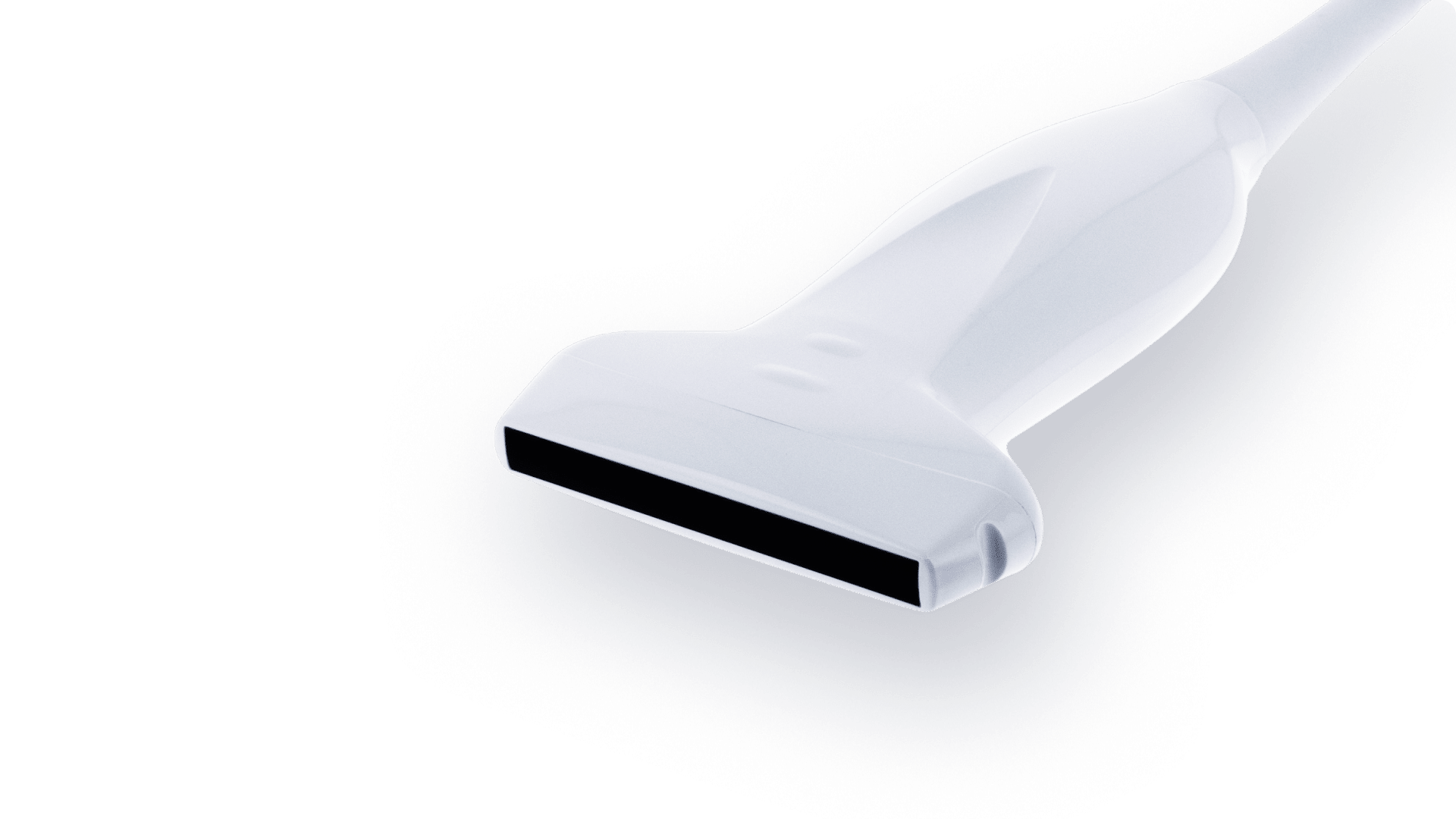
Linear Array – 8.5MHz – 192 elts
LA 8.5/192
-
Center Frequency 8.5
-
Number elements 192
-
Pitch 0.2
-
Radius curvature N/A
-
Transverse aperture 4
-
Focal Depth 20
Linear Array – 8.0MHz – 192 elts
LA 8.0/192
-
Center Frequency 8.0
-
Number elements 192
-
Pitch 0.286
-
Radius curvature N/A
-
Transverse aperture 4
-
Focal Depth 20