

Radiology
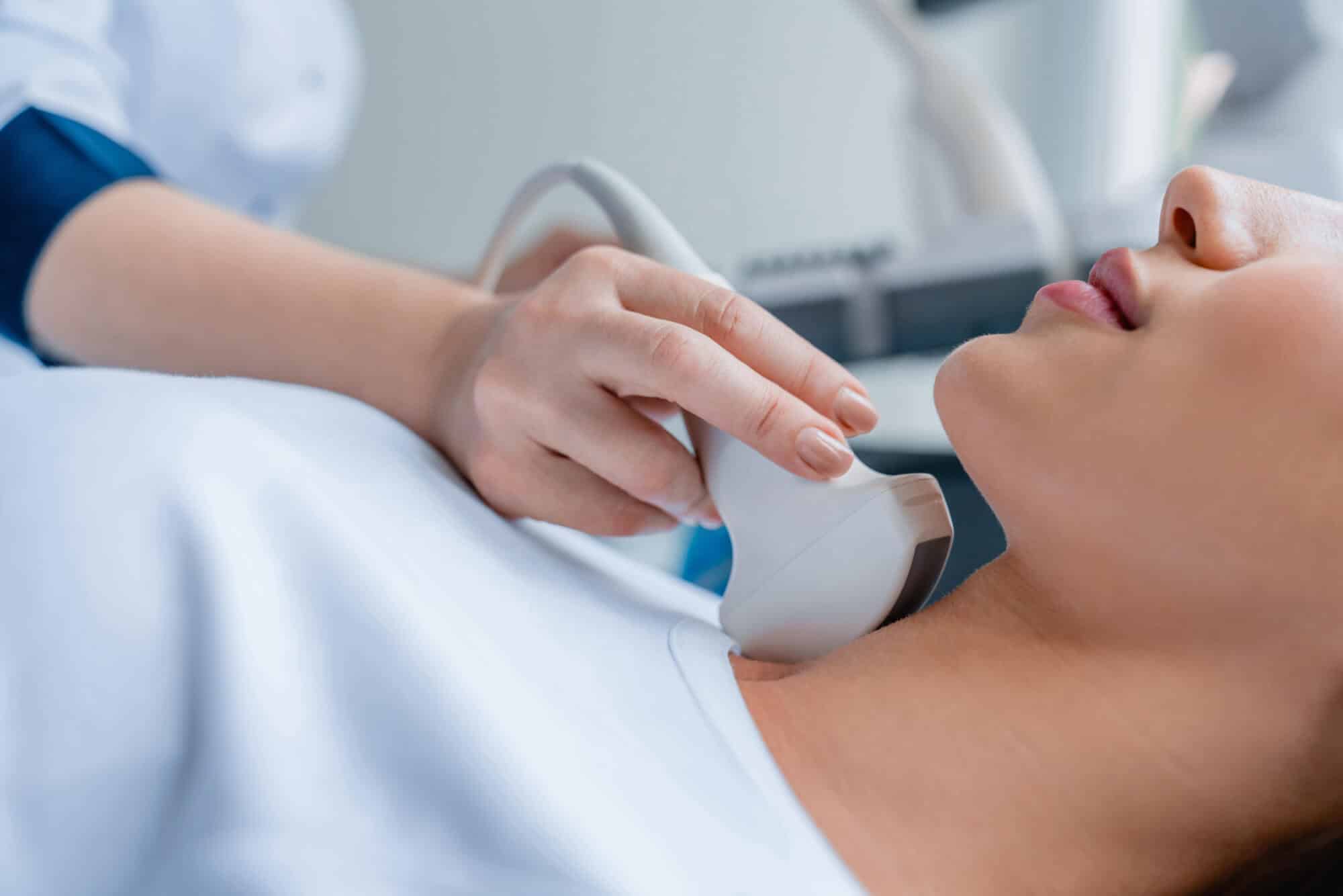
In the radiology field, ultrasound imaging stands out as a cutting-edge modality. It empowers clinicians with real-time, non-invasive visualization of a wide variety of soft tissue structures. Its precision and availability aid in a quick diagnosis establishment of a broad range of conditions, improving patient care by providing accurate insights.
Thyroid
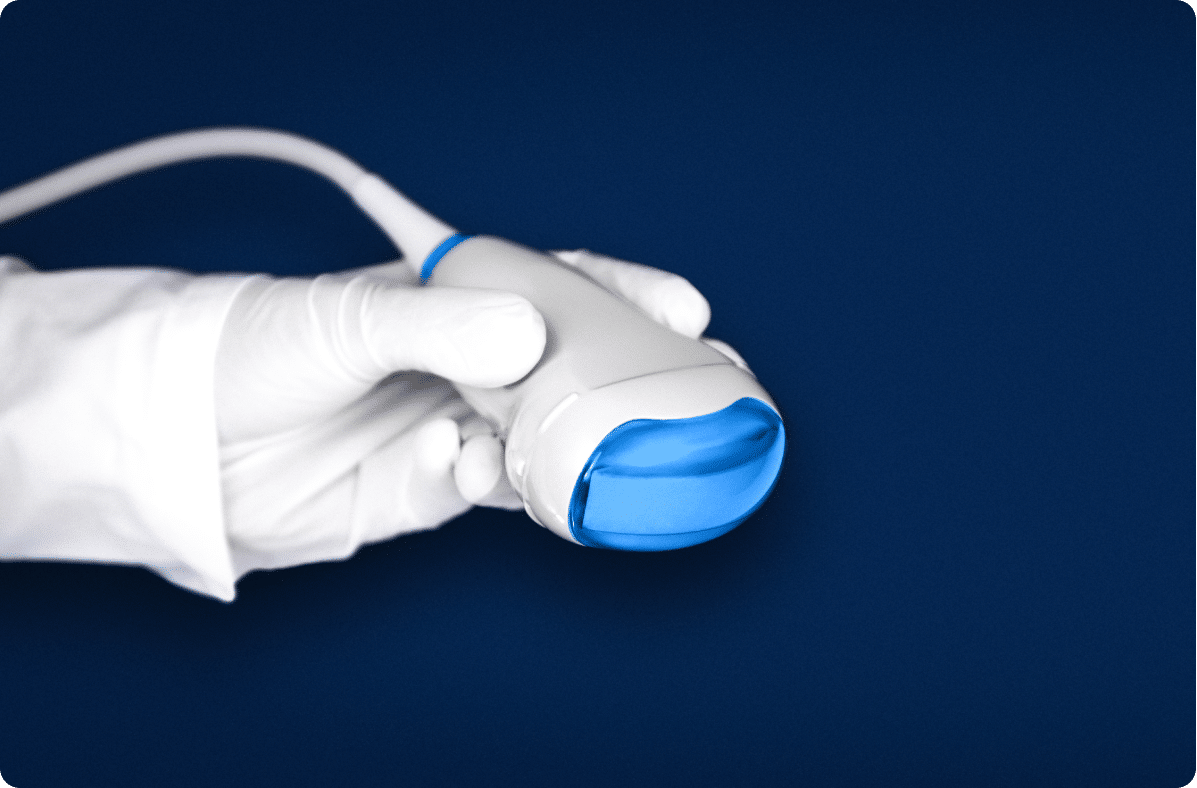
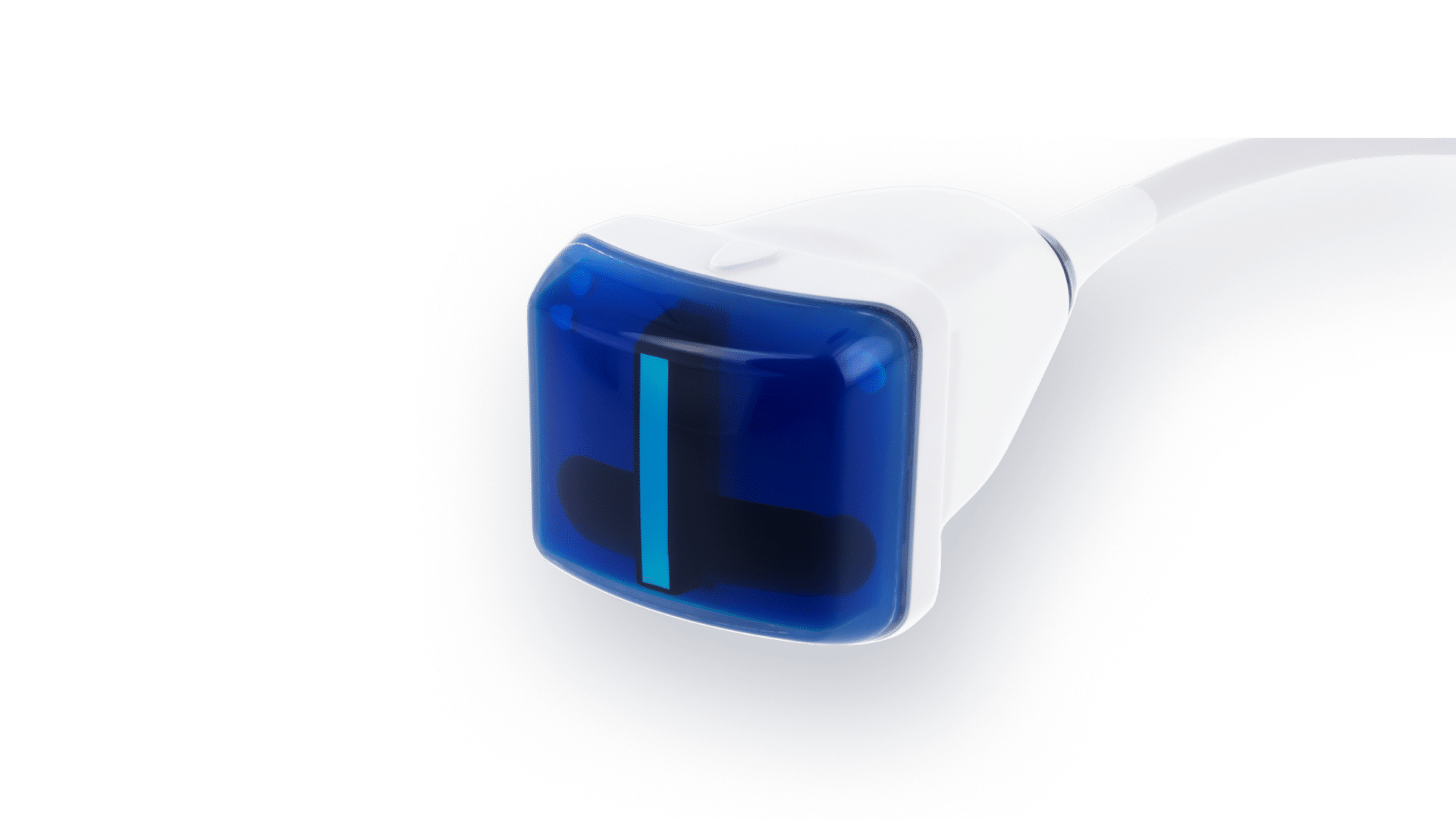
The thyroid, a superficial and soft structure located in the neck, is readily examined with a linear array probe, recommended to operate at central frequencies from 8.0 to 10.0 MHz.
Thyroid Ultrasound is the most appropriate imaging modality to distinguish normal thyroid parenchyma from diffuse (hypothyroidism, hyperthyroidism, and thyroiditis) or nodular thyroid disease (hyperplastic thyroid gland, adenomas or thyroid cancer).
A comprehensive evaluation in gray-scale ultrasound of consistency, echogenicity, margins and invasion of the surrounding structures in the neck is essential. It may be assisted by color Doppler ultrasound to evaluate the nature of the overall blood perfusion and the stage of a potential disease.
Accurate measurements of thyroid lobe diameter and the localization of focal lesions are also crucial components of this clinical evaluation.
1
Thyroid
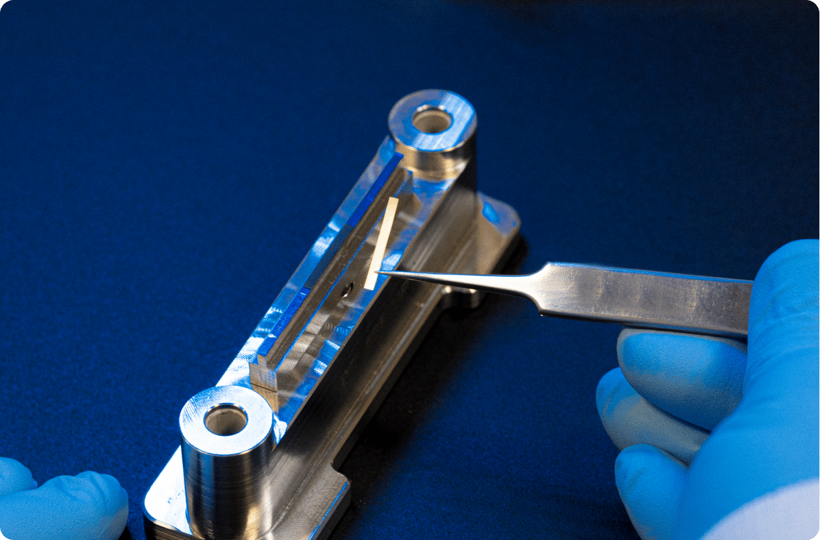
Recommanded probes :
Linear Array – 8.5MHz – 256 elts
LA 8.5/256
-

Center Frequency 8.5
-

Number elements 256
-

Pitch 0.2
-

Radius curvature N/A
-

Transverse aperture 4
-

Focal Depth 20
Linear Array – 10.0MHz – 256 elts
LA 10.0/256
-
Center Frequency 10.0
-
Number elements 256
-
Pitch 0.15
-
Radius curvature N/A
-
Transverse aperture 3
-
Focal Depth 13
Breast
Breast Ultrasound is usually carried out with a linear array probe, typically operating at central frequencies from 8.5 MHz to 14.0 MHz.
Integrating clinical palpation and pathological assessment with ultrasound allows clinicians to effectively characterize all breast tissue types.
The Breast Imaging Reporting And Data System (BIRADS) is a required standardized lexicon to describe and classify findings and their probability of malignancy.
This approach provides detailed insights related to lesions morphology and vascularization, enhancing diagnosis accuracy and guiding appropriate treatment decisions.
A significant cancer risk factor is dense breast tissue and suspicious findings include irregular shape; indistinct and spiculated margins; acoustic shadowing; hypoechoic echotexture; and calcifications.
Complementary volume scanning, employing a motor-driven linear array probe, enables full-field volume acquisition, reducing operator errors, enhancing lesion contour precision and facilitating therapy follow-up.
2
Breast
Recommanded probes :
Linear Array 3D Wobble – 8.5MHz – 192 elts
LA 8.5/192-4D
-
Center Frequency 8.5
-
Number elements 192
-
Pitch 0.2
-
Radius curvature N/A
-
Transverse aperture 4
-
Focal Depth 20
Linear Array – 8.0MHz – 192 elts
LA 8.0/192
-
Center Frequency 8.0
-
Number elements 192
-
Pitch 0.286
-
Radius curvature N/A
-
Transverse aperture 4
-
Focal Depth 20
Liver
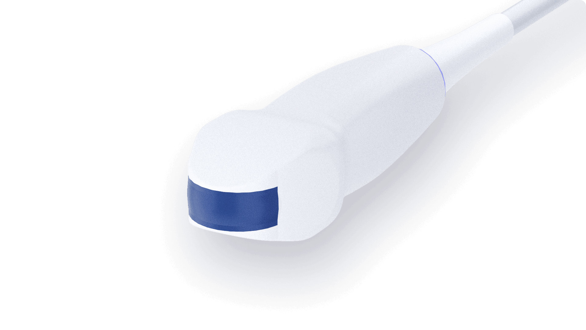
The liver is easily accessible using a curved array probe, typically operating at central frequencies from 3.0 MHz to 6.5 MHz.
Liver ultrasound is commonly recommended for fatty food intolerance, abnormal liver functions tests and non-obstructive jaundice.
The healthy echotexture of the liver can be assessed by its homogeneity and mid-grey tones, otherwise when a brighter live echogenicity is visible, it can suggest a pathological condition, such as cirrhosis or fatty infiltration.
3
Liver
Recommanded probes :

Curved Array – 3.5MHz – 192 elts
CLA 3.5/R60/192-SiX-Boost
-
Center Frequency 3.5
-
Number elements 192
-
Pitch 0.332
-
Radius curvature 60
-
Transverse aperture 11
-
Focal Depth 65
Curved Array – 3.5MHz – 192 elts
CLA 3.5/R60/192-SiX
-
Center Frequency 3.5
-
Number elements 192
-
Pitch 0.332
-
Radius curvature 60
-
Transverse aperture 11
-
Focal Depth 65
Lungs
The lungs are imaged using a small footprint probe for accessing between the ribs ideally operating at at central frequencies from 2.5 MHz to 6.5 MHz for optimal depth penetration.
During the last decade sonography was proven useful for assessing conditions where fluid or masses accumulate in the pleura (thin membrane surrounding the lungs).
It also helps detect lung anatomical changes resulting from respiratory viruses, including SARS-CoV-2.
4
Lungs
Recommanded probes :
Microconvex Array – 6.5MHz – 128 elts
CLA 6.5/R11.5/128-Microconvex
-
Center Frequency 6.5
-
Number elements 128
-
Pitch 0.216
-
Radius curvature 11.5
-
Transverse aperture 6
-
Focal Depth 35
Microconvex Array – 6.5MHz – 192 elts
CLA 6.5/R11.5/192-Microconvex
-
Center Frequency 6.5
-
Number elements 192
-
Pitch 0.144
-
Radius curvature 11.5
-
Transverse aperture 6
-
Focal Depth 35
Kidneys
Renal examination is usually carried out with a curved array probe, typically operating at central frequencies from 3.5 MHz to 5.0 MHz.
For the initial evaluation of renal insufficiency, ultrasound is the preferred screening modality.
Patients are positioned with arms raised above the head to optimize visualization of the kidneys by elevating the lower costal margin and widening the intercostal spaces.
Chronic renal diseases are detected by smaller, fibrotic kidneys that appear relatively brighter with thinner parenchyma.
Ultrasound is highly sensitive and specific in identifying obstructive renal conditions like renal calculi;, complex cysts; solid masses; renal vein thrombosis; and hydronephrosis related to UTIs (Urinary Tract Infections).
Additionally, ultrasound is instrumental in monitoring post-lithotripsy recovery, post-transplantation follow-up, and in guiding biopsies to evaluate for transplant rejection.
5
Kidneys
Recommanded probes :
Curved Array – 3.5MHz – 192 elts
CLA 3.5/R60/192-SiX-Boost
-
Center Frequency 3.5
-
Number elements 192
-
Pitch 0.332
-
Radius curvature 60
-
Transverse aperture 11
-
Focal Depth 65
Curved Array – 5.0MHz – 192 elts
CLA 5.0/R40/192
-
Center Frequency 5.0
-
Number elements 192
-
Pitch 0.285
-
Radius curvature 40
-
Transverse aperture 11
-
Focal Depth 50