Obstetrics & Gynecology
This medical field encompasses two distinct sub-specialties. Obstetrics (OB) is dedicated to managing pregnancy, childbirth, and the postpartum period. In contrast, Gynecology (GYN) concentrates on the examination and treatment of the female reproductive system.
Ultrasound is the preferred imaging modality for assessment of pregnancy and female pelvis in real-time due to the absence of related risks to reproductive organs and fetuses.

Foetus / Uterus
For early pregnancy ultrasound examinations, transvaginal endocavity probes at around 7.5 MHz are employed to evaluate the fine structures of the developing embryo. As the pregnancy progresses, transabdominal ultrasound becomes the standard for fetal assessment, using typically a curved array probe at around 5.0 MHz.
Obstetric ultrasound revolutionized the care of the pregnant woman, considered as the method of choice for screening, diagnosis, intervention and monitoring pregnancy.
In the early stages of pregnancy, as early as 5 to 6 weeks, transvaginal endocavity probes allow the evaluation of the developing embryo.
For the following pregnancy stages, transabdominal real-time ultrasound is the routine method for fetal assessment.
In addition, volume imaging improves the visualization of facial, extremities and structural anomalies.
1
Foetus / Uterus
Recommanded probes :

Endocavity Curved Array Probe – 6.5MHz – 192 elts
CLA 6.5/R11.5/192
-
Center Frequency 6.5
-
Number elements 192
-
Pitch 0.144
-
Radius curvature 11.5
-
Transverse aperture 6
-
Focal Depth 35

Curved Array 3D Wobble – 5.0MHz – 192 elts
CLA 5.0/R40/192-4D
-
Center Frequency 5.0
-
Number elements 192
-
Pitch 0.285
-
Radius curvature 40
-
Transverse aperture 11
-
Focal Depth 60
Ovaries / Uterus / Cervix
The most commonly used imaging method for female pelvic examination employs transabdominal imaging with curved array probe from 3.5 MHz to 5.0 MHz and transvaginal endocavity probe at around 7.5 MHz, preferred for precise gynecological diagnostics due to its proximity to pelvic organs.
Gynecological ultrasound investigations are conducted for women presenting symptoms such as palpable pelvic masses, menstrual irregularities, postmenopausal bleeding, and pelvic pain.
Alterations in the uterine structure’s size, shape, contour, and echotexture, as revealed by ultrasound, may indicate conditions like myomas or adenomyosis, which can impact pregnancy outcomes and fertility.
Hystero-salpingo Contrast Sonography (HyCoSy) is a dynamic, transvaginal ultrasound procedure used in early infertility assessments. It evaluates uterine anomalies and checks for blockages or damage in the fallopian tubes that could negatively affect fertilization and implantation, employing specialized contrast agents for clearer imaging.
2
Ovaries / Uterus / Cervix
Recommanded probes :

Endocavity Curved Array Probe – 7.5MHz – 192 elts
CLA 7.5/R8.8/192
-
Center Frequency 7.5
-
Number elements 192
-
Pitch 0.144
-
Radius curvature 8.8
-
Transverse aperture 6
-
Focal Depth 35
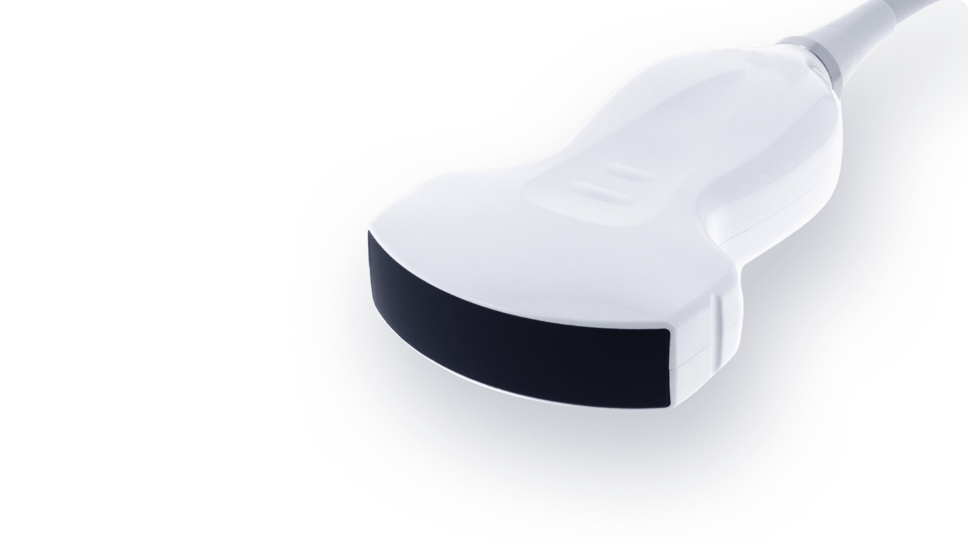
Curved Array – 3.5MHz – 192 elts
CLA 3.5/R60/192-SiX-Boost
-
Center Frequency 3.5
-
Number elements 192
-
Pitch 0.332
-
Radius curvature 60
-
Transverse aperture 11
-
Focal Depth 65
